b value mri
Materials and Methods Thirty-four consecutive patients with vertebral compression fractures underwent sagittal diffusion-weighted imaging DWI with different b values. Objective The objective was to explore the optimal b value in diffusion-weighted imaging DWI of MRI for differential diagnosis of benign and malignant vertebral fractures.

Epos Trade
Depending on the organ being imaged b-values typically range from 50-1000smm 2.
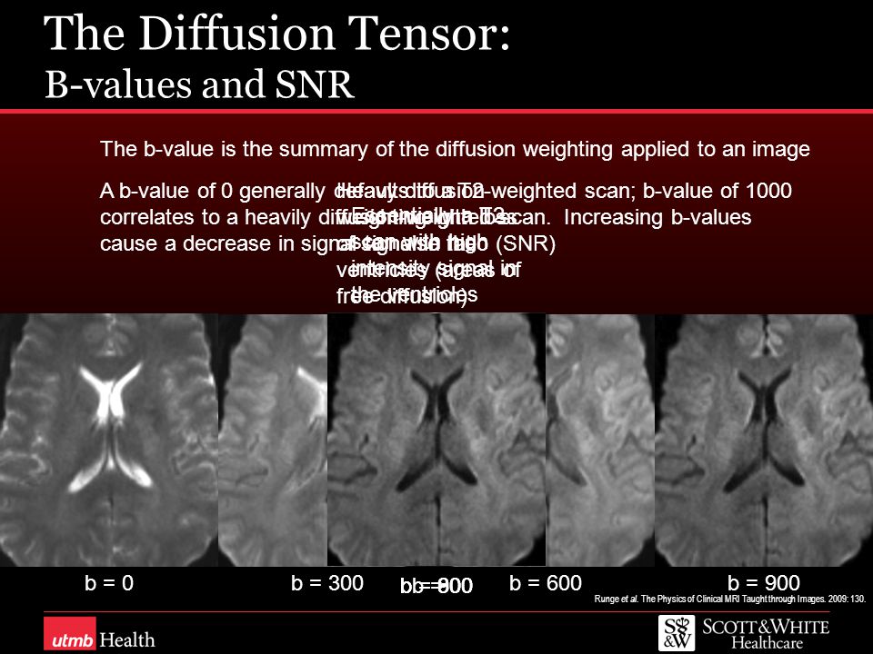
. Forty-two consecutive patients underwent multi b-value 16 evenly spaced b-values between 0 and 2000 smm 2 DWI along with multi-parametric MRI MP-MRI of the prostate at 3 Tesla followed by trans-rectal ultrasoundMRI fusion guided targeted biopsy of suspicious lesions detected at MP-MRI. In general approximately 1000 smm 2 is the maximal b value for DWI 5 11. The b factor summarizes the influence of the gradients on the diffusion weighted images.
Lesions were analyzed for benignitymalignity using apparent diffusion coefficient ADC values with 10 b-value combinations and by measuring the lesionnormal parenchyma ADC ratio. Studies have reported that the use of b values higher than 1000 smm 2 and 2000 smm 2 improves tumor localization and the contrast between benign and malignant lesions in the prostate and the breasts 12 14. The use of b values more than 1000 smm 2 would offer better contrast but was more liable to suffer susceptibility artifact.
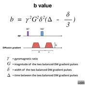
Thus image sequences from low to high b-values must be read side by side in order to establish the precise anatomic location of the suspected tumor. B value measures the degree of diffusion weighting applied thereby indicating the amplitude G time of applied gradients δ and duration between the paired gradients Δ and is calculated as. The degree of diffusion weight in g correlates with the strength of the diffusion gradients characterized b y the b - value which is a function of the gradient related parameters.
The reason is readily apparent from the images below. Computed DWI images up to a simulated b-value of. Mike Notohamiprodjo Die Radiologie Munich Germany Optimization and Longterm Monitoring in Radiology Departments 14.
Some recent studies have proposed that imaging with very high b-values. Mean ADC values were approximately 13 1-15 higher in. Numerous studies have shown that restricted diffusion is the single most reliable predictor of cancer on multi-parametric MRI.
Large b factors should be chosen for more precise evaluation of ADC values of the tumor. Using lower b-values such as 0 and 50 together with higher b-values 600 smm2 was beneficial Az0928 and 0927. Vikas Gulani University of Michigan Ann Arbor MI USA Improving Productivity in MRI Clinical Experience in a Multi-site Outpatient Practice Setting 1.
There is no consensus on the optimal b-value. Either of these conditions lead to a higher signal loss for a given rate of diffusion in a region in the body. Suppose we acquire two images - one without applying these GMFs b 0 and then another with a certain b value eg b 1000.
The Value of MR Imaging. A b value of 8001000 smm 2 would provide an excellent spatial resolution and an adequate signalnoise ratio for lesion evaluation. Certa in illnesses show restrictions of diffusion for example demyel in ization and cytotoxic edema.
A baseline b-value of 50 smm² is often used in liver diffusion-weighted imaging instead of b 0. Generally the larger the b values used the lower the ADC values owing to the contribution of perfusion. At b 3000 smm 2 points from 1 to 3 were assigned to tumor signals the same as those observed at b 1000 smm 2 but 4 points were assigned to hyperintense signals between those of the normal subcortical white matter and the corticospinal tract and 5 to markedly hyperintense signals that were equal to or higher than that of the normal.
Modern scanners usually allow a range of 0-4000smm 2. Our purpose was to evaluate the appearance of the normal brain on DW MR images as the diffusion gradient strength b value is increased from 1000 to 3000 smm2. MRI Resources MR Myelography - Diffusion Weighted Imaging - DICOM - Calculation - Universities - Contrast Agents B-Value The b-value is a factor of diffusion weighted sequences.
As MR scanner hardware has improved allowing for increased gradient strengths we are able to generate higher b values for diffusion-weighted DW imaging. Routine abdominal MRI and DWI were performed using seven b-values 0 50 200 400 600 800 1000 smm 2. Mean ADC values were significantly different between malignant and benign lesions for all b-value combinations P0000.
The higher the value b the stronger the diffusion weighting. Therefore a larger b value is achieved by increasing the gradient amplitude and duration and by widening the interval between paired gradient pulses. The best b-value combination was 0 and 800 Az0935.
Diffusion-weighted images are obtained by acquiring T2-weighted images with the addition of diffusion weighting gradient known as the b value. With b 0 bright signals are noted in multiple veins due to the high T2 of blood coupled with sluggish flow. Strength duration and the period b etween diffusion gradients.
A higher b value would mean that the GMFs are either applied for longer or that their magnitude is higher.

High B Value Diffusion Weighted Mr Imaging Of Normal Brain At 3t European Journal Of Radiology

Combining Perfusion And High B Value Diffusion Mri To Inform Prognosis And Predict Failure Patterns In Glioblastoma International Journal Of Radiation Oncology Biology Physics

Illustration Of Dw Mri Signal Decay Of Various Types Of Tissues With Download Scientific Diagram

Diffusion Weighted Imaging Radiology Reference Article Radiopaedia Org

Diffusion Mri Signal Decay Versus B Value A The Diffusion Signal Download Scientific Diagram

Illustration Of The Signal Decrease When The Echo Time And The B Value Download Scientific Diagram

Figure 3 From Zafer Koc Value For Characterization Of Liver Lesions B Diffusion Weighted Mri And Optimal Semantic Scholar

High B Value Diffusion Weighted Mr Imaging Of Adult Brain Image Contrast And Apparent Diffusion Coefficient Map Features American Journal Of Neuroradiology
Diffusion Weighted Imaging Radiology Reference Article Radiopaedia Org

Physical Principles Use Of High B Values And Clinical Applications Of Diffusion Weighted Imaging Of Ischemic Stroke

Advantage Of High B Value Diffusion Weighted Imaging For Differentiation Of Common Pediatric Brain Tumors In Posterior Fossa European Journal Of Radiology
How Does B Value Affect Hardi Reconstruction Using Clinical Diffusion Mri Data Plos One

Conventional Mri Dwi And Adc Images Of Braf V600e Mutant And Download Scientific Diagram

Zafer Koc Value For Characterization Of Liver Lesions B Diffusion Weighted Mri And Optimal Semantic Scholar
How Does B Value Affect Hardi Reconstruction Using Clinical Diffusion Mri Data Plos One

Fig 2 High B Value Diffusion Weighted Mr Imaging Of Suspected Brain Infarction American Journal Of Neuroradiology

Impact Of B Value On Estimates Of Apparent Fibre Density Biorxiv
Technical Considerations In Brain Dwi Ppt Video Online Download

Signal Intensity Changes With Increasing B Values A 29 Year Old Female Download Scientific Diagram


Comments
Post a Comment